
Extracellular micro vesicles and exosomes and their role in the insect cell-baculovirus expression system
SUPERVISOR: REINGARD GRABHERR
Background.
Besides direct cell-to-cell contact, eukaryotic cells communicate through various extra-cellular vesicles (Ratajczak et al., 2006). In principle, two types of vesicles exist: (i) micro vesicles that are budding from the plasma membrane and (ii) exosomes that are released by exocytosis of multivesicular endosomes (Cocucci et al., 2009). Micro vesicles are spherical with a diameter of up to 1 µm, while exosomes are in the range of 40-100 nm. Intensive exosome research has been focusing on various mammalian cell types, e.g. epithelial cells (van Niel et al., 2007), neurons (Faure et al., 2006) and cancer cells (Wolfers et al., 2001). However, exosomes and extracellular vesicles have also been discovered in Caenorhabditis elegans (Ligeois et al., 2006), Leishmania parasites (Silverman et al., 2010) and Drosophila cells (Koppen et al., 2011).
Based on the finding that extracellular vesicles confer messages between cells by transporting specific proteins, nucleic acids, and lipids, it becomes clear that understanding this type of cell-to-cell communication is highly important for research in cell biology. In addition, there are technological aspects to exosomes and microvesicles, as they are also present in cell-based production processes. Yet, little is known to date about their impact on the performance of a biotechnological production process, the product yield and quality. Recently, it was shown that enrichment of exosomes in the culture medium during the production process leads to inhibition of apoptosis of CHO cells (Han and Rhee, 2018). Also, exosomes are often part of a biopharmaceutical product, e.g. when secreted virus-like particles (VLPs) or whole virus vaccines are being manufactured and thus, have a yet unknown influence on product quality.
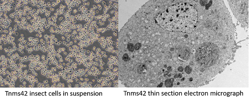
For the production of complex proteins, one attractive production platform is the insect cell/baculovirus expression system, due to easy genetic manipulation and high yield production. In the past, we have expressed complex proteins and multi subunit particles in Spodoptera frugiperda and Trichoplusia ni insect cell lines (Krammer et al., 2010; Palmberger et al., 2012; Klausberger et al., 2013; Wilde et al., 2014; Nika et al., 2017). The insect cell/baculovirus system is a viral expression system and we have shown that host cells react not only to the infection by baculoviruses but also to the metabolic burden caused by recombinant protein production. Transcriptome analyses identified several genes specifically up-regulated when complex secreted proteins are being produced. (Koczka et al., 2018). The impact on production and composition of exosomes and extracellular vesicles in this process has yet not been considered. However, the question arises, whether such vesicles transmit signals to neighboring cells, influence susceptibility to infection or whether and how they reflect the cells’ stress during viral infection and recombinant protein production (as does the transcriptome).
Aims and methods.
The goal of this project is to evaluate the role of exosomes and microvesicles during baculovirus infection of insect cells and their relevance during a protein production process. Especially for continuous processes reproducible and constant culture conditions are highly important, and incomplete process control has often hampered large-scale production of biopharmaceuticals in baculovirus-infected insect cells.
Therefore, we are planning to isolate all extracellular vesicles by density gradient centrifugation and to characterize these vesicles in terms of their proteome, lipid composition as well as any potentially contained nucleic acids. So far, no analytical methods exist. Hence, based on these results, we will establish tools for the detection and quantification of vesicles (e.g. by specific antibodies) in insect cells. In parallel we will do transcriptomics analysis. Different insect cell lines will be tested, either uninfected, infected by an empty baculovirus or infected by a baculovirus encoding a recombinant protein. Previously, we have analyzed the changes in the transcriptional pattern for these different states (Kozcka et al., 2018) and found quite a few differences, depending if and what kind of recombinant protein was being expressed (secreted or non-secreted protein).
In this new study the secreted influenza A virus hemagglutinin and the intracellular yellow fluorescent protein will serve as model proteins. By analyzing the distribution and types of extracellular vesicles, we will learn whether they play a role as stress markers and whether they can be used as a process control parameter. The newly generated proteome data will be compared to transcriptome data. Based on these analyses, genes that are up-regulated under certain conditions and code for proteins that abundantly end up in extracellular vesicles will be identified. Further the effect of enrichment of exosomes and/or microvesicles as well as their depletion will be investigated. The final goal is to understand vesicle-based communication between insect cells and to generate new tools for process monitoring as well as process control.
Collaborations within this thesis include GRILLARI (RAMAN measurements for lipid composition), ALTMANN (Proteomics) and JUNGBAUER (separation of vesicles).
Cocucci, E., Racchetti, G., Meldolesi, J. (2009) Shedding microvesicles: artefacts no more. Trends Cell. Biol. 19, 43-51. doi: 10.1016/j.tcb.2008.11.003
Faure, J., Lachenal, G., Court, M., Hirrlinger, J. (2006) Exosomes are released by cultured cortical neurons. Mol. Cell. Neurosci. 31, 642-648. doi: 10.1016/j.mcn.2005.12.003
Han, S., Rhee W.J. (2018) Inhibition of apoptosis using exosomes in Chinese hamster ovary cell culture. Biotechnol. Bioeng. 115, 1331-1339. doi: 10.1002/bit.26549
Klausberger, M., Wilde, M., Palmberger, D., Hai, R., Albrecht, R. A., Margine, I., Hirsh, A., Grabherr, R., Krammer, F. (2013) One-shot vaccination with an insect cell-derived low-dose influenza A H7 virus-like particle preparation protects mice against challenge. Vaccine 32, 355-362. doi: 10.1016/j.vaccine.2013.11.036.
Koczka, K., Peters, P., Ernst, W., Himmelbauer, H., Nika, L., Grabherr, R. (2018) Comparative transcriptome analysis of a Trichoplusia ni cell line reveals distinct host responses to intracellular and secreted protein products expressed by recombinant baculoviruses J. Biotechnol. 270, 61–69. doi: 10.1016/j.jbiotec.2018.02.001
Koppen, T., Weckmann, A., Müller, S., Staubach, S., Bloch, W., Dohmen, R. J., Schwientek, T. (2011) Proteomics analyses of microvesicles released by Drosophila Kc167 and S2 cells. Proteomics 11, 4397-4410. doi: 10.1002/pmic.201000774
Krammer, F., Schinko, T., Messner, P., Palmberger, D., Ferko, B., Grabherr, R. (2010) Influenza virus-like particles as antigen-carrier platform for an ESAT-6 epitope of Mycobacterium tuberculosis. J. Virol. Methods. 167, 17-22. doi: 10.1016/ j.jviromet.2010.03.003
Ligeois, S., Benedetto, A., Garnier, J. M., Schwab, Y. Labouesse, M. (2006) The V0-ATPase mediates apical secretion of exosomes containing Hedgehog related proteins in Caenorhabditis elegans. J. Cell. Biol. 173, 949-961. doi: 10.1083/jcb.200511072
Nika, L., Wallner, J., Palmberger, D., Koczka, K., Vorauer-Uhl, K., Grabherr R. (2017) Expression of full-length HER2 protein in Sf9 insect cells and its presentation on the surface of budded virus-like particles. Protein Expr. Purif. 136, 27-38. doi: 10.1016/j.pep.2017.06.005
Palmberger, D., Wilson, I.B.H., Berger, I., Grabherr, R., Rendic, D. (2012) SweetBac: A new approach for the production of mammalianised glycoproteins in insect cells. PloS One 7:e34226. doi: 10.1371/journal.pone.0034226
Ratajczak, J., Wysoczynski, M., Hayek, F., Janowska-Wieczorek, A., Ratajczak, M.Z. (2006) Membrane derived microvesicles: important and underappreciated mediators of cell-to-cell communication. Leukemia 20, 1487-1495. doi: 10.1038/sj.leu.2404296
Silverman, J.M., Clos, J., de Oliveira, C.C., Shirvani, O., Fang, Y., Wang, C., Foster, L.J., Reiner, N.E. (2010) An exosome-based secretion pathway is responsible for protein export from Leishmania and communication with macrophages. J. Cell Sci. 123, 842-852. doi: 10.1242/jcs.056465
Van Niel, G., Raposo, G., Candalh, C., Boussac, M., Hershberg, R., Cerf-Bensussan, N., Heyman, M. (2007) Intestinal epithelial cells secrete exosome-like vesicles. Gastroenterology 2001, 337-349. doi: 10.1053/gast.2001.26263
Wilde, M., Klausberger, M., Palmberger, D., Ernst, W., Grabherr, R. (2014) Tnao38, High Five and Sf9 - Evaluation of host virus interactions in three different insect cell lines: baculovirus production and recombinant protein expression. Biotechnol. Lett. 36, 743-749. doi: 10.1007/s10529-013-1429-6
Wolfers, J., Lozier, A., Raposo, G., Regnault, A. Théry, C., Masurier, C., Flament, C., Pouzieux, S., Faure, F., Tursz, T., Angevin, E., Amigorena, S., Zitvogel, L. (2001) Tumor- derived exosomes are a source of shared tumor rejection antigens for CTL cross-priming. Nat. Med. 7, 297-303. doi: 10.1038/85438