
Deciphering the folding and inhibition mechanism of the staphylococcal myeloperoxidase inhibitor SPIN
SUPERVISOR: Christian OBINGER
Background.
The heme peroxidase myeloperoxidase (MPO), one of five human peroxidases, is a key player in neutrophil-mediated killing of phagocytosed pathogens. Its’ main mechanism of action is the hydrogen peroxide-mediated oxidation of halides and pseudo-halides, primarily chloride and thiocyanate. The resulting highly reactive oxidants indiscriminately attack and kill invading bacteria with high efficiency (Kettle et al. 1997, Nauseef et al. 2014). However, this unspecific innate immune response comes with a price at inflammatory conditions when MPO is released from the phagosome into blood. Aberrant MPO activity has been associated with chronic inflammatory diseases including atherosclerosis, neurodegenerative disease, lung disease, arthritis, and kidney disease, and there is mounting evidence for its detrimental role in tumour progression (Davies et al. 2008). The development of small molecule inhibitors, however, is significantly hampered by MPO’s high structural similarity to the other human peroxidases, where inhibition due to off-target effects would have severe consequences. Homologous peroxidases are thyroid peroxidase, which play crucial roles in hormone biosynthesis in the thyroid gland (Ruff et al. 2005) and peroxidasin, which is involved in the formation of basement membranes by crosslinking of collagen IV (Péterfi et al 2014). To date no inhibitors are available for clinical application of MPO (Malle et al. 2007).
However, the ingenuity of nature has provided a potential new solution in the form of a small protein evolved by Staphylococcus aureus, one of only a handful of pathogens able to escape phagocytosis (Spaan et al. 2013). This protein, called SPIN (Staphylococcal Peroxidase Inhibitor), is an eight kDa protein consisting of a C-terminal MPO-binding domain and an N-terminal inhibitory peptide, which inserts into the substrate channel and active site of MPO in a currently unknown mechanism (de Jong et al 2017 and 2018). The aim of this project is to understand the kinetics and thermodynamics of SPIN binding to and inhibition of MPO. A special focus will be placed on the mechanism of folding of the N-terminal peptide, which – after binding of the C-terminal domain to MPO - mediates formation of the final inhibitory MPO-SPIN complex with picomolar affinity (Leitgeb et al. 2022).
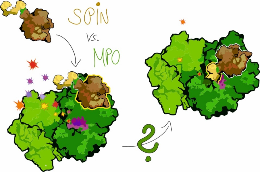
Aims and methods.
SPIN variants will be generated in order to elucidate the sequence of SPIN binding via the SPIN C-terminal domain, folding of the SPIN N-terminal domain and formation of the final inhibitory SPIN-MPO complex. Based on the recently solved crystal structure (Leitgeb et al. 2022), site-directed mutagenesis of selected residues will be performed to elucidate the role of key amino acids in folding, binding and inhibition. SPIN variants with increased affinity and specificity towards MPO will be further engineered by directed evolution and the best candidate will be selected based on thermal and protease stability, inhibitory capacity and selectivity towards MPO with respect to other human peroxidases. Binding to and inhibition of MPO by wild-type SPIN-aureus and the generated variants will be characterized by a broad set of biochemical/biophysical methods including (i) X-ray crystallography including anomalous data collection for identifying substrates in the substrate binding site, (ii) detailed kinetic and thermodynamic analyses of the generated variants and their interaction with MPO by complimentary techniques, e.g. sequential-mixing stopped-flow spectroscopy with UV-vis, CD- and fluorescence detectors, differential scanning calorimetry, isothermal titration calorimetry, surface plasmon resonance spectroscopy, (iii) MD simulations, and (iv) investigation of inhibitory action(s) in biological assays. Together, these approaches will provide the basis to generate the first truly specific protein-based inhibitor of myeloperoxidase without side effects.
Collaborations.
Collaborations within BioToP and BOKU: Chris OOSTENBRINK (MD simulations) and Core Facilities Biomolecular and Cellular Analyses as well as Mass Spectrometry.
External collaboration: Pierre VAN ANTWERPEN (Universite Libre de Bruxelles, Laboratory of Pharmaceutical Chemistry & Analytical Platform, Brussels, Belgium) (cell-based MPO activity assays).
Kettle, A. J., Winterbourn, C. C., (1997) Redox Rep, 3, (1), 3-15, DOI: 10.1080/13510002.1997.11747085
Nauseef, W. M., Borregaard, N., (2014) Neutrophils at work. Nature Immunology, 15 (7), 602-611, DOI: 10.1038/ni.2921
Davies, M. J., Hawkin C. L., Pattison, D. I., Rees, M. D. (2008). Antioxid Redox Signal. 10 (7), 1199-234, DOI: 10.1089/ars.2007.1927
Ruff, j., Carayon, P., (2005) Arch Biochem Biophys., 445 (2). 269-77, DOI: 10.1016/j.abb.2005.06.023
Péterfi, Z., Geiszt, M., (2014). Trends Biochem Sci. 39 (7), 305-7, DOI: 10.1016/j.tibs.2014.05.005
Malle, E., Furtmuller, P. G., Sattler, W., Obinger, C., (2007) Br J Pharmacol, 152 (6), 838-54, DOI: 10.1038/sj.bjp.0707358
Spaan, A. N., Surewaard, B. G., Nijland, R., van Strijp, J. A., (2013) Annu Rev Microbiol, 67, (1), 629-50, DOI: 10.1146/annurev-micro-092412-155746
de Jong, N. W. M., Ramyar, K. X., Guerra, F. E., Nijland, R., Fevre, C., Voyich, J. M., McCarthy, A. J., Garcia, B. L., van Kessel, K. P. M., van Strijp, J. A. G., Geisbrecht, B. V., Haas, P. A., (2017). Proc Natl Acad Sci U S A 2017, 114, (35), DOI: 9439-9444, 10.1073/pnas.1707032114.
de Jong, N. W. M., Ploscariu, N. T., Ramyar, K. X., Garcia, B. L., Herrera, A. I., Prakash, O., Katz, B. B., Leidal, K. G., Nauseef, W. M., van Kessel, K. P. M., van Strijp, J. A. G., Geisbrecht, B. V., (2018). J Biol Chem 2018, 293, (7), 2260-2271, DOI: 10.1074/jbc.RA117.000134.