
Partitioning of virus-like particles and protein superstructures in charged hydrogels
SUPERVISOR: ALOIS JUNGBAUER
Background.
Virus-like particles (VLP) are one of the most important candidates for modern vaccines and drug delivery. They are non-pathogenic, can be manufactured for a wide range of proteins and exert sufficient stability to be formulated as drugs. Purification of VLP is still a complex process, and usually gradient ultracentrifugation, filtration, or conventional size exclusion chromatography are used. These processes are slow and have a low productivity.
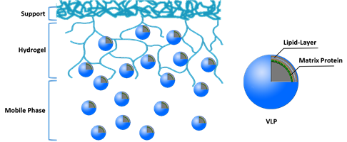
Charged hydrogel or chromatography beads in form of polymer-grafted media have a very high protein binding capacity (Jungbauer, 2005). It has been observed that they also bind large biomolecules such as plasmids and viruses although these cannot penetrate into the pores of the chromatography material. We hypothesize that these large molecules bind at the outer surface and partition into the outer shell of the beads. The large molecules or particles never reach the centre of the beads. Binding at the outer layer is sufficient to provide sufficient binding capacity. The 10% radius of the outer shell of a sphere makes up 1/3 of the volume. It is not clear yet why these large molecules can penetrate the outer layer.
The high protein binding capacity of charged hydrogels has been explained by solid diffusion. A charged hydrogel is quickly saturated by proteins and this cannot be explained by pore diffusion, since the pores are too small in such hydrogels. The protein is partitioning into the hydrogel and then is diffusing along the fibres of the gel. Although the diffusivity is very low the driving force for the fast mass transfer is the concentration gradient. For proteins up to 150 kDa this mechanism is well understood, while it is not understood for larger biomolecules such as plasmids and VLP. For pDNA, having a similar size as VLP and protein superstructures, we have postulated that adsorption is completely entropy driven (Tarmann and Jungbauer, 2008). The plasmid molecules arrange in a vertical manner in a kind of self-assembly process. Yet, it is not possible to extrapolate from plasmids to VLP, large proteins or protein superstructures. Plasmids are highly uniformin their charge, and very flexible, while the adiabatic compressibility of proteins and VLP seems to be very low. They are very compact molecules and when they arrange at a surface they are not able to change their conformation. For viruses embedded into continuous media such as monoliths we have observed that the adsorption is driven only by convention and adsorption (Gerster et al., 2013)., while diffusional phenomena do not play a major role.
Aims and methods.
The aim of the project is to elucidate how VLP, large proteins and protein superstructures partition into highly charged hydrogels and polymer-grafted media. We hypothesize that this is affected by solid diffusion and by assembly of these proteins and particles into a uniform layer on the surface of the beads.
As model proteins IgG, thyroglobulin, IgM and von Willebrand factor (vWF), influenza virus-like particles produced in insect cells, and HIV virus-like particles produced in CHO cells will be used (STEINKELLNER, GRABHERR). vWF with a molecular mass of 2000 kDa is a typical representative of a protein superstructure. IgG, thyroglobulin and IgM wil be used in order to understand the effect of size. Proteins will range from 150 to 2000 kDa, and the VLP diameter range is from 100-200 nm, so an entire size range from 10 nm (IgG) to 200 nm (HIV VLP) will be covered. For studying charged hydrogels a polymer grafted acrylamido material and an agarose based material with dextran grafts will be used. For control experiments non-grafted media will be used. The uptake will be studied with conventional batch uptake experiments using a batch contactor. Only pseudo-equilibrium isotherms can be determined because we expect that within the time of investigation equilibrium will not be achieved. Attainment of equilibrium would require incubation for several weeks and the material would probably deteriorate beforehand. The uptake will also be followed microscopically using confocal laser microscopy and TEM (SCHAEFFER). The layer thickness will be determined over time. In order to understand the arrangement of the large molecules and VLP the adsorption will be also studied in isothermal titration calorimetry (ITC) and flow calorimetry (OBINGER). Flow calorimetry for high loadings and ITC for low loadings will be used if changes in the binding mechanism are affected by the density of the molecules on the surface. The heat of adsorption will be measured and the enthalpy of the interaction determined. Adsorption kinetics will also be made at different temperatures with measurement of bothentropy and enthalpy. The difference of calorimetric and van′t Hoff analysis will be used to get insight if a conformational change is taking place during adsorption. To corroborate this, SAXS analysis will be performed with loaded and unloaded chromatography media, and information on the 3D structure will be obtained from the fractal dimension, radius of gyration and a coarse grained model (Tscheliessnig et al., 2013). The derived structure will be then compared with the structure determined in solution (Mueller, 2013).
Gerster, P., Kopecky, E.M., Hammerschmidt, N., Klausberger, M., Krammer, F., Grabherr, R., Mersich, C., Urbas, L., Kramberger, P., Paril, T., Schreiner, M., Noebauer, K., Razzazi-Fazeli, E., Jungbauer, A. (2013) Purification of infective baculoviruses by monoliths. J. Chromatogr.A, 1290, 36-45
Jungbauer, A. (2005) Chromatographic media for bioseparation. J. Chromatogr. A 1065, 3-12
Mueller, M., Loh, M.Q.T., Tscheliessnig, R., Tee, D.H.Y., Tan, E., Bardor, M., Jungbauer, A. (2013) Liquid formulations for stabilizing IgMs during physical stress and long-term storage. Pharm. Res., 30, 735-750
Tarmann, C., Jungbauer, A. (2008) Adsorption of plasmid DNA on anion exchange chromatography media. J. Sep. Sci. 31, 2605-2618
Tscheliessnig, R., Zoernig, M., Herzig, E.M., Lueckerath, K., Altrichter, J., Kemter, K., Paunel-Goerguelue, A., Loegters, T., Windolf, J., Pabisch, S., Cinatl, J., Rabenau, H.F., Jungbauer, A., Mueller-Buschbaum, P., Scholz, M., Koch, J. (2012) Nano-coating protects biofunctional materials. Materials Today 15, 394-404