
Biochemistry of firmicute coproporphyrin ferrochelatase from Listeria monocytogenes
SUPERVISOR: Christian OBINGER
Background.
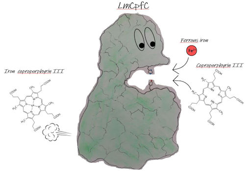
Prokaryotic heme b biosynthesis has been studied extensively in the past. While most diderm (Gram-negative) bacteria synthesize the iron containing cofactor analogously to mammals via the so-called "protoporphyrin-dependent" (PPD) pathway, monoderm (Gram-positive) organisms utilize a different pathway, the "coproporphyrin-dependent" (CPD) route (Dailey et al., 2015). In the CPD as well as in the PPD pathway a ferrochelatase is needed to incorporate ferrous iron into the porphyrin ring. Importantly, the respective porphyrin substrate is different in each pathway. Coproporphyrin III has two more propionate groups than protoporphyrin IX. These two additional propionates potentially have a major impact to substrate-enzyme interactions and other structural properties translating to functional aspects. Until 2015 all coproporphyrin ferrochelatases of monoderm bacteria were studied in the believe to be protoporphyrin ferrochelatases and consequently all biochemical and biophysical studies were performed with a non-physiological substrate. In this project we aim at understanding of this recently identified CPD heme biosynthesis pathway. The essentiality of heme for monoderm bacteria (Mayfield et al., 2013) and the differences in heme biosynthesis between monoderm bacteria and humans, make all enzymes of this pathway promising targets for the development of novel antibacterial therapeutic substances.
Coproporphyrin ferrochelatases (CpfCs) have only been marginally studied with the correct substrate (Hobbs et al., 2017, Hofbauer et al., 2019). First kinetic and structural data of the wild-type enzyme will be a perfect basis for further structural and functional studies of CpfC variants, in which residues essential for catalysis and for substrate binding and stabilization will be altered. Also, the interaction with other proteins, delivering the ferrous iron or producing the other substrate, coproporphyrin III, or receiving the product, coproheme, will be focus of research.
Aims and methods.
Monomeric CpfC from Listeria monocytogenes (wild-type and variants) will be heterologously expressed in E. coli. Based on recently solved X-ray structures from apo- and coproheme-bound Lm CpfC (Hofbauer et al., 2019) site-directed mutagenesis of distal and proximal amino acids will be performed. Emphasis will be placed on amino acids involved in coproporphyrin III and coproheme binding and residues involved in coordinating incoming ferrous iron and other regulatory sites.
Wild-type and mutant proteins will be characterized by a broad set of biochemical/physical methods including (i) X-ray crystallography, (ii) detailed spectral analysis (UV-Vis, electron paramagnetic resonance and resonance Raman spectroscopy) of proteins, and protein-based radicals in different redox- and spin-states (in cooperation with Giulietta SMULEVICH, Department of Chemistry, University of Florence, Italy), (iii) time-resolved multi-mixing UV-Vis stopped-flow studies in order to analyze the kinetics of substrate binding and iron insertion, (iv) spectroelectrochemical studies (in cooperation with Gianantonio BATTISTUZZI from the Department of Chemistry, University of Modena and Reggio Emilia, Italy). These methods will provide valuable insight into the mechanism of ferrous iron inseration.
REFERENCES:
Dailey, H. A., Gerdes, S., Dailey, T. A., Burch, J. S. & Phillips, J. D. (2015). Proc Natl Acad Sci U S A 112, 2210-2215.
Hobbs, C., Reid, J. D. & Shepherd, M. (2017).Biochem J 474, 3513-3522.
Hofbauer, S., Helm, J., Obinger, C., Djinović-Carugo, K. & Furtmüller, P. G. (2020). FEBS J., in press
Mayfield, J. A., Hammer, N. D., Kurker, R. C., Chen, T. K., Ojha, S., Skaar, E. P. & DuBois, J. L. (2013).J Biol Chem 288, 23488-23504.