
Biochemical and Molecular Analysis of the Cell Wall Glyco(prote)ome of Lactobacillus buchneri
SUPERVISOR: PAUL MESSNER
Project assigned to: EVA SMOLAR
Background.
Lactobacilli belong to the lactic acid bacteria (LAB) and are one of the industrially most important groups of bacteria (Kleerebezem et al., 2010), which play a key role in industrial and artisan food raw-material fermentation, including a large variety of fermented dairy products. Due to their GRAS status, Lactobacillus buchneri is an ideal candidate organism for cell surface display of biofunctional compounds.
The cell wall of lactobacilli is composed of different macromolecules together determining the strain-specific properties of the organisms. A thick multilayered PG (peptidoglycan) layer can be decorated with teichoic acids (WTA-wall teichoic acid and/or LTA-lipoteichoic acid), proteins and EPS (exopolysaccharide). SDPs (sortase-dependent proteins) and S-layers are well studied in lactobacilli, but many other types of cell surface proteins and protein anchors exist and have not been investigated.
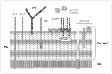
Usually the S-layer protein is attached to the underlying peptidoglycan sacculus via charged or uncharged secondary cell wall polymers (Mesnage et al., 2000; Sara, 2001; Schäffer & Messner, 2005). However, in LABs, the SCWPs (secondary cell wall polymers) are only poorly characterized. Therefore, especially the identification of the S-layer anchoring system to the cell wall will have a significant impact on the application potential of this organism, such as its development as an in vivo cell surface display system (Åvall-Jääskeläinen et al., 2002; Desvaux et al., 2006; Zarschler et al., 2010a).
Aims and methods.
Since the whole genome sequence of L. buchneri CD034 is available it will be data-mined for the occurrence of glycosylation-related loci that could be involved in the biosynthesis of the diferrent cell wall components. Further, the molecular analysis of the involved genes in the biosynthesis of these molecules will be examined.
In several bacilli "non-classical" SCWPs (Schäffer & Messner, 2005) have been identified as anchor-ing structure between S-layer and peptidoglycan (Mesnage et al., 2000; Sara, 2001; Leoff et al., 2008). We expect that the possible oligosaccharide structure of the anchor molecule is different from that of the S-layer glycoprotein glycan. To get first hints about the presence of this anchor molecule, the native peptidoglycan/S-layer complex, comprising the anchor, will be isolated. Because of the expected non-covalent linkage between the anchor molecule and the S layer protein, separation can be achieved by using high-resolution gel-filtration chromatography and RP-HPLC. In addition, the structural and molecular characterization of the S-layer anchoring system of L. buchneri CD034 will be performed. Binding studies using the isolated anchor compound and purified S-layer glycoprotein will be carried out to confirm the proposed function of the anchor molecule. By interfering with its biosynthesis the binding of the S-layer glycoprotein to the anchor molecule should be abolished.
In the past it has never been investigated whether S-layer-carrying LABs are able to synthesize one of the mentioned exocellular polymers (EPS). If EPS will be found on L. buchneri CD034, attempts will be undertaken to abolish its biosynthesis by insertional inactivation of key genes. Eventually it will help to identify possible cross reactions with other glycosylated cell wall compounds and thus results in a better overall understanding of the cell wall glyc(oprote)ome of L. buchneri CD034.
Åvall-Jääskeläinen, S., U. Hynönen, N. Ilk, D. Pum, U. B. Sleytr, and A. Palva. 2008. Identification and characterization of domains responsible for self-assembly and cell wall binding of the surface layer protein of Lactobacillus brevis ATCC 8287. BMC Microbiology 8:165.
Kleerebezem, M., P. Hols, E. Bernard, T. Rolain, M. Zhou, R.J. Siezen, and P.A. Bron. 2010. The extracellular biology of the lactobacilli. FEMS Microbiol. Rev. 34:199-230.
Leoff, C., B. Choudhury, E. Saile, C.P. Quinn, R.W. Carlson, and E.L. Kannenberg. 2008. Structural elucidation of the nonclassical secondary cell wall polysaccharide from Bacillus cereus ATCC 10987. Comparison with the polysaccharides from Bacillus anthracis and B. cereus type strain ATCC 14579 reveals both unique and common structural features. J. Biol. Chem. 283:29812-29821.
Mesnage, S., T. Fontaine, T. Mignot, M. Delepierre, M. Mock, and A. Fouet. 2000. Bacterial SLH domain proteins are non-covalently anchored to the cell surface via a conserved mechanism involving wall polysaccharide pyruvylation. EMBO J. 19:4473-4484.
Sara, M. 2001. Conserved anchoring mechanisms between crystalline cell surface S-layer proteins and secondary cell wall polymers in Gram-positive bacteria. Trends Microbiol. 9:47-49.
Schäffer, C. and P. Messner. 2005. The structure of secondary cell wall polymers: how Gram-positive bacteria stick their cell walls together. Microbiology 151:643-651.
Zarschler, K., B. Janesch, B. Kainz, R. Ristl, P. Messner, and C. Schäffer. 2010a. Cell surface display of chimeric glycoproteins via the S-layer of Paenibacillus alvei. Carbohydr. Res. 345:1422-1431.